Blood in the urine, a raised PSA, a testicular lump or an unexpected scan finding does not establish cancer on its own, but each can justify a structured specialist assessment. By the end of the consultation, you should understand which organ is being investigated, which tests answer the remaining questions, whether tissue is needed, and when treatment decisions can be made.
Key takeaways
- Bring previous scans, pathology reports, medication lists, and symptom dates.
- Expect questions about bleeding, pain, urinary changes, weight loss, and family history.
- Ask why each test is needed, when results will arrive, and what happens next.
- Request the biopsy site, pathology diagnosis, grade, stage, and treatment options in writing.
When should you book the consultation, and what should you bring?
Book a genitourinary cancer consultation in Jaipur for visible blood in urine, a testicular lump or enlargement, persistent flank pain, unexplained weight loss, urinary obstruction, or pain that does not settle. An abnormal PSA, urine cytology, kidney-function result, ultrasound, CT, MRI, or biopsy also warrants review.
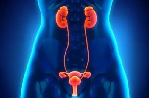
A urologic cancer specialist assesses cancers of the prostate, bladder, kidney, testis, penis, ureter, renal pelvis and adrenal gland.
- Bring original digital files for ultrasound, CT, MRI or PET-CT, not only printed reports.
- Carry biopsy reports, pathology slides or paraffin blocks, laboratory results, PSA records, operation notes, and discharge summaries.
- Write a symptom timeline, including blood in urine, fever, pain, weight change, urinary difficulty and testicular or genital changes.
- List every medicine, supplement and allergy, and note smoking, prior cancers and family history.
- Ask who will review the pathology, what stage is suspected, which alternatives are reasonable, and how delay affects outcomes.
| Finding | Usual pathway | When another referral matters |
|---|---|---|
| Visible blood in urine | Urology assessment with upper-tract imaging and possible cystoscopy | A negative dipstick or cytology does not cancel evaluation |
| Female bladder, ureter or urethral mass | Urologic cancer specialist | Refer to gynecologic oncology if it suggests ovarian, cervical, uterine, vaginal or vulvar cancer, or pelvic cancer invading the urinary tract |
| Testicular lump | Urology assessment with ultrasound and tumour markers | Do not arrange a scrotal needle biopsy before specialist review |
What happens during registration, history-taking and examination?
Registration records your identity, contact details and referral reason, then the genitourinary cancer doctor in Jaipur reviews why you came and what has already been found. Describe when each symptom began, whether it is worsening, and any blood in urine, pain, urinary change, lump or weight loss.
Bring the original digital scan files, not only printed reports.
The examination is focused on the suspected organ and your symptoms:
- Abdomen: checks for tenderness, swelling or an enlarged kidney, liver or mass.
- Flanks: assesses pain, fullness or kidney-area tenderness.
- Genitals: looks for penile or scrotal skin changes, swelling and other abnormalities.
- Testicles: compares both sides for a lump, firmness, size change or tenderness.
- Lymph nodes: checks regions such as the groin for enlargement.
- Digital rectal examination: assesses the prostate when prostate symptoms, an abnormal PSA or another finding makes it relevant.
The doctor also records every medicine, supplement, allergy and previous operation, along with smoking history and cancers in close relatives. Review may include blood and urine results, ultrasound, CT, MRI or PET-CT images, biopsy reports, pathology slides or blocks.
Ask whether outside pathology needs specialist review; details such as Gleason Grade Group, bladder muscle invasion, tumour type and grade can change the diagnosis or treatment plan. Expect the visit to clarify the next test or review, not necessarily provide a final stage that day.
How does the doctor decide which tests you need?
The doctor matches your symptom pattern, examination and existing results before ordering tests. Blood in the urine raises concern for bladder, kidney or upper-tract cancer, but infection, stones and prostate conditions can cause it too; a negative dipstick or urine cytology does not cancel evaluation of high-risk haematuria.
A urology cancer consultation in Jaipur may therefore include cystoscopy and upper-tract imaging.
The test choice usually follows this pattern:
| Test | What it helps distinguish | When it applies |
|---|---|---|
| Urine culture and cytology | Infection versus abnormal urinary cells | Burning, fever, visible blood or persistent haematuria |
| Renal-function tests | Whether the kidneys work safely and contrast can be used | Before CT urography, contrast CT or treatment |
| PSA and prostate examination | Prostate enlargement, prostatitis or possible prostate cancer | Raised PSA or prostate abnormality; PSA alone is not a diagnosis |
| Ultrasound | Stones, cysts, blockage, prostate enlargement or a testicular mass | Initial assessment of urinary, kidney or scrotal symptoms |
| Multiparametric MRI | Suspicious prostate areas and biopsy targets | Suspected clinically localized prostate cancer |
| CT urography | Kidneys, ureters and bladder lining from the upper tract | Haematuria or suspected urothelial tumour |
| Cystoscopy | Direct view of the bladder and urethra | Persistent or high-risk haematuria |
| Biopsy and staging scans | Tissue diagnosis and spread to lymph nodes, lungs or bones | When imaging identifies a suspicious lesion |
A renal mass is not automatically cancer, and a suspicious MRI is not proof. The specialist may recommend surveillance, repeat imaging or biopsy, while staging scans follow confirmed or strongly suspected cancer.
What does a biopsy and pathology discussion involve?
Tissue is required for a definitive cancer diagnosis because scans show shape and spread, not the cells themselves. A suspicious multiparametric MRI can identify a prostate target and guide the decision to biopsy, but it cannot confirm cancer or assign its grade.
Biopsy planning depends on the organ involved:
- Prostate: a targeted, systematic or combined biopsy obtains cores through the perineum or rectum after MRI review.
- Bladder: transurethral resection removes visible tumour and samples the bladder wall; the report must state whether detrusor muscle is present and invaded.
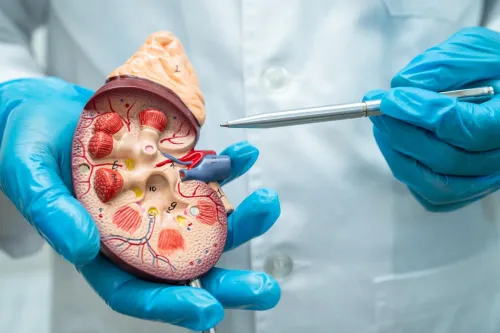
- Kidney: an image-guided core biopsy is planned when tissue will change management, such as distinguishing a renal tumour from lymphoma or metastasis.
- Testis: a solid testicular mass is usually assessed with bilateral scrotal ultrasound, followed by serum AFP, beta-hCG and LDH before treatment.
Doctors usually avoid a needle biopsy through the scrotum for a suspected testicular tumour because it can disrupt tissue planes and affect the definitive surgical approach. Normal AFP, beta-hCG and LDH do not exclude cancer.
The pathology discussion separates diagnosis, grade and stage. Ask whether the report identifies seminoma or nonseminoma, renal-cell subtype, tumour type, grade, margins and other microscopic features. In prostate cancer, Gleason Grade Group describes aggressiveness; in bladder cancer, muscle invasion changes the treatment pathway.
TNM stage often requires imaging, and an expert review of outside slides or blocks can change classification or treatment planning with a urologic cancer specialist in Jaipur.
What happens after the consultation, and what should you ask before leaving?
A diagnosis is not the same as a stage. Expect a separate results appointment to review the pathology, imaging and laboratory findings, assign TNM stage, and discuss treatment choices.
Outside slides or paraffin blocks may be sent for specialist pathology review; details such as Gleason Grade Group, bladder muscle invasion, seminoma versus nonseminoma, tumour grade and renal-cell subtype can change the plan.
- Ask which organ is involved, what tumour type and grade the report shows, and what TNM stage is suspected.
- Ask whether outside pathology needs review before surgery, chemotherapy, radiotherapy or active surveillance, who will review it, and when you will receive the interpretation.
- Ask how each option could affect urine control, urinary diversion, erections, ejaculation, testosterone, sensation and daily activity.
- If treatment could damage fertility, ask about sperm cryopreservation before treatment. Request prompt andrology referral; testosterone or fertility supplements do not replace cancer treatment, and testosterone can suppress sperm production.
- Ask who coordinates follow-up, which doctor receives scan and blood-test results, the next appointment date, warning symptoms, and how urgently treatment must begin.
At Jaipur AndroUrology Center, ask how the team reviews results with you, records the stage, and turns the findings into a documented next-step plan. A genitourinary cancer clinic in Jaipur should also explain which decisions require a multidisciplinary referral and give you a contact route for unanswered questions.
Frequently asked questions
When should you book a genitourinary cancer consultation in Jaipur?
Book one for visible blood in urine, a testicular lump or enlargement, persistent flank pain, unexplained weight loss, urinary obstruction, or pain that does not settle. Arrange review of an abnormal PSA, urine cytology, kidney-function result, ultrasound, CT, MRI, or biopsy.
What should you bring to a genitourinary cancer consultation?
Bring previous urine and blood tests, PSA results, ultrasound or scan images and reports, biopsy slides or pathology reports, a medication list with doses, allergy details, and a short timeline of symptoms. Include relevant family cancer history.
How does a doctor decide which tests are needed?
The doctor combines your symptoms, examination findings, age, medical history, kidney function, and existing reports. The next test depends on the suspected organ and question, such as imaging for a mass, urine testing for urinary-tract findings, or biopsy for tissue diagnosis.
What does a biopsy and pathology discussion involve?
The doctor explains where tissue will be taken, how the procedure is performed, possible risks, and when the report is expected. Ask for the exact pathology diagnosis, tumour type, grade, stage or extent, and whether further tests are needed.
What should you ask before leaving the consultation?
Ask what the working diagnosis is, which tests come next, how to prepare, when and how results will be shared, which symptoms need urgent care, and when the follow-up appointment will occur.
Keywords